Personalized Medical Care & Dedicated International Patient Concierge

Medical Guide
What Is Re-Root Canal Treatment, and When Is It Needed?
Re-root canal treatment is a second root canal on an already-treated tooth. It is needed when symptoms or imaging show infection is still in the canal.
Schedule a ConsultationWhat Is Re-Root Canal Treatment, and When Is It Needed?
Medically reviewed by Dr. Park Hyo Jin, Specialist in Conservative Dentistry / Director
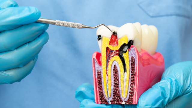
Re-root canal treatment is a second root canal on a tooth that has already had one. Dentists call it endodontic retreatment: the old filling material is taken out of the roots, the canal system is cleaned again, and the space is resealed. It is needed when a previously treated tooth shows evidence that infection is still present or has come back — tenderness on biting, gum swelling, a draining spot on the gum, or a dark area at the root tip on an X-ray that has not shrunk or has grown. It is not needed simply because a tooth has an old root canal in it, and it is not the answer when the discomfort is coming from somewhere else.
What "Re-Root Canal Treatment" Means in Plain Terms
Re-root canal treatment means redoing the inside of a tooth that was already root-canal treated, working through the crown of the tooth rather than through the gum. You may see it written several ways — endodontic retreatment, nonsurgical retreatment, root canal revision, second root canal treatment — and they all describe the same thing. Endodontics is the branch of dentistry concerned with the soft tissue inside a tooth and the space it occupies.
The American Association of Endodontists describes retreatment as a second chance to save a tooth that healed improperly or became painful or diseased months or even years after the first procedure. That framing matters, because patients often assume a failed root canal means a lost tooth.
It also helps to be clear about what it is not: not gum surgery at the root tip, not an extraction, not an implant or a bridge, and not simply a new crown — though a new restoration is usually needed afterward.
This article stays on one question: what re-root canal treatment is, and when a dentist decides it is needed. For the appointment itself — what is done step by step and what published outcome data shows for retreated teeth — see the companion article in this library, Re-Root Canal Treatment: Can a Failed Root Canal Be Saved?
Why a Sealed Root Canal Can Become Infected Again
A root canal can fail because the inside of a tooth is a branching space that cannot be perfectly sterilized, and because the seal on top of it does not last forever. The American Association of Endodontists groups post-treatment endodontic disease into four possible causes: microorganisms retained or reintroduced inside the canal system, microorganisms surviving in the tissues just outside the root tip, foreign body reactions in those tissues, and true cysts at the root end. Only the first is treated by re-root canal treatment; the other three are addressed surgically if at all. That single fact explains much of when retreatment is offered and when it is not.
Anatomy that was never found or never cleaned
Root canals are rarely the simple straight tubes drawn in diagrams. A pooled analysis of cone beam computed tomography studies found a second canal in the largest root of the upper first molar — the canal endodontists call MB2 — in most of those teeth, with detection rates varying sharply according to the method used to look. That is the practical point: a canal that exists is not always a canal that was seen. Fine side branches, connecting channels between canals, and sharply curved ends can all hold tissue and bacteria the first treatment never reached. None of this necessarily means the first dentist made an error.
Bacteria that survived inside the tooth
Where infection persists, it is usually because bacteria remained in the canal system or found their way back in. A review in the International Endodontic Journal by Siqueira concluded that most endodontic failures result from persistent or secondary infection inside the root, with infection outside the root implicated in a smaller share of cases. Bacteria in a treated canal often live in a biofilm — a layered community stuck to the canal wall — which resists cleaning solutions far better than free-floating organisms, so a canal can look adequately filled on an X-ray and still hold a pocket of it in a recess instruments never touched.
Leakage from above
The seal on top of a root canal is part of the treatment, not an afterthought. A study of endodontically treated teeth in the International Endodontic Journal by Ray and Trope found the technical quality of the coronal restoration strongly associated with the health of tissue at the root tip, mattering at least as much as the root filling itself. Saliva carries bacteria, and saliva reaching the canal through a gap re-contaminates it. Common routes include:
- A crown or filling that has loosened, chipped, or come off.
- New decay at the margin of an existing crown.
- A long delay between finishing the root canal and placing the permanent restoration.
A crack or a root fracture
A crack changes the question entirely, because it creates a route for bacteria that no filling material can close. A vertical root fracture often produces few symptoms at first and may be recognized only when the surrounding bone breaks down in a distinctive pattern that three-dimensional imaging can reveal.
What "Failure" Actually Means in Clinical Language
A "failed" root canal usually means the tissue around the root tip is still inflamed, not that the tooth has collapsed. Dentists record two diagnoses for every tooth: one for the pulp inside and one for the tissues around the root. A tooth that has had a root canal is recorded as previously treated, and the second diagnosis describes what the surrounding tissue is doing. Three categories come up constantly:
- Symptomatic apical periodontitis — inflammation around the root tip causing pain on biting, tapping, or pressure over the root, with or without a visible change on the X-ray.
- Asymptomatic apical periodontitis — the same inflammation and bone destruction seen as a dark area on an X-ray, with no symptoms at all.
- Chronic apical abscess — a low-grade infection with gradual onset, little or no discomfort, and intermittent discharge of pus through a small channel in the gum.
A tooth carrying one of those diagnoses has post-treatment disease, a more accurate phrase than "failure" because it names something specific and treatable. Equally important is what does not count: a treated tooth that is tender for a while and then settles has not failed, and a dark area smaller than it was on an earlier film is healing.
Symptoms That Suggest a Treated Tooth Needs Another Look
The usual signal is discomfort in a tooth you had stopped thinking about, and there is a reason it can still hurt. Root canal treatment removes the nerve tissue inside the tooth, but the ligament suspending the root in bone keeps its own nerve supply and registers pressure. So a treated tooth cannot feel hot or cold, yet it can feel distinctly sore when you bite or when the bone around the root is inflamed. What patients most often describe:
- Tenderness when biting on that specific tooth, or a sense that it sits higher than its neighbors.
- A dull pressure or ache that comes and goes over weeks rather than a sharp jolt.
- Swelling in the gum near the root, which may rise and fall.
- A small bump on the gum that drains and reappears, sometimes with a salty or foul taste.
- Discomfort returning after a long symptom-free stretch, occasionally years later.
Then there is the category that produces nothing to report. Asymptomatic apical periodontitis is defined by the absence of symptoms and shows up as a dark area on a radiograph taken for another reason. The absence of pain is not by itself evidence that a root canal succeeded.
What the Imaging Actually Shows
Dental images show bone and tooth structure, not bacteria, so a dentist reads them for the footprint infection leaves behind. Around a healthy treated root, the thin white line of bone looks continuous and the dark ligament space looks even. Where inflammation is active, that ligament space widens and bone at the root tip loses density, appearing as a darker zone. Two limits are worth knowing: a standard X-ray is a flat picture of a three-dimensional object, so a lesion on the cheek side of a root can hide behind the root; and one image is a snapshot, while two images years apart show direction.
For teeth with post-treatment disease, the American Association of Endodontists and the American Academy of Oral and Maxillofacial Radiology state in a joint position statement that limited field of view cone beam computed tomography — a small-volume three-dimensional dental scan — is the imaging method of choice for evaluating non-healing after previous endodontic treatment. Such imaging can identify untreated canals, root perforations, the quality of the previous filling, and bone loss patterns suggesting a fracture. In studies cited by the association, adding three-dimensional data to otherwise complete diagnostic information changed the treatment plan in 35 to 62 percent of cases. Magok Pureunmaeum Dental Clinic lists 3D CT imaging among its facility capabilities, and one cone beam CT unit is publicly listed as available there. Whether a scan is warranted in your case is a clinical judgment, not a routine step.
How a Dentist Confirms the Diagnosis
No single test decides this; the diagnosis is assembled from several findings that must agree. The American Association of Endodontists compares endodontic diagnosis to a jigsaw puzzle, with a probable diagnosis formed from the history and then confirmed by examination and testing. A retreatment assessment usually includes:
- History — when the original root canal was done, what was placed afterward, and how the current symptoms behave.
- Soft-tissue examination — swelling, and any sinus tract, the small opening in the gum through which a chronic abscess drains.
- Periodontal probing — a single deep, narrow pocket in an otherwise healthy gum is a warning sign of a root fracture.
- Percussion, palpation, and a bite test — tapping the tooth, pressing the gum over the root, and biting cusp by cusp on a small plastic instrument, always compared against nearby healthy teeth.
- Pulp testing of the neighbors — the treated tooth will not respond to cold, so adjacent teeth are tested to confirm they are not the true source.
- Radiographs — at least two views of the treated tooth from different angles, plus any older images that can be obtained.
- Sinus tract tracing — a small flexible point threaded into a draining spot and imaged, so the channel leads back to the root actually responsible.
One principle deserves repeating: treatment should not be provided without a diagnosis. If the tests conflict, the appropriate next step may be to reassess later, rather than to open a tooth and hope.
Conditions That Can Imitate a Failed Root Canal
Pain near a treated tooth does not prove the treated tooth is the problem. Nerve fibers from different structures converge before reaching the brain, so the place you point to is not always the place that hurts. A systematic review of nonodontogenic toothache — tooth pain arising from something other than a tooth — grouped the causes into categories including muscle pain and sinus pain referred to the teeth, nerve-related pain, and pain with no identifiable dental origin. Conditions commonly mistaken for a failing root canal include:
- A neighboring tooth with an inflamed nerve. Pain from an untreated tooth often refers along the jaw, and patients frequently name the wrong tooth.
- A crack in a different tooth. A cracked cusp typically produces sharp pain on release of biting pressure rather than on pressure itself.
- Gum and bone infection. A periodontal abscess arises from a deep pocket around the root, not from inside the canal, and is treated differently.
- Bite overload. A restoration sitting slightly high, or heavy clenching, can inflame the ligament and make a tooth sore to bite on with no infection involved.
- Sinus inflammation. Because upper back tooth roots sit close to the sinus floor, congestion can present as pressure in several upper molars at once.
- Jaw muscle and joint disorders. The National Institute of Dental and Craniofacial Research describes temporomandibular disorders as a group of more than 30 conditions affecting the jaw joint and chewing muscles, and their pain can be felt in the teeth.
Getting this wrong is not neutral: retreating a tooth that was never the source removes tooth structure and leaves the real cause unaddressed.
When Re-Root Canal Treatment Is Not Indicated
Retreatment is not indicated when the tooth is healing normally, when the trouble is not inside the canal, or when the tooth cannot be restored afterward. A responsible plan is as specific about these situations as about the cases that call for treatment.
- The tooth is still healing. Bone regenerates slowly, and a dark area measurably smaller than on an earlier image is a healing lesion.
- The tooth is comfortable and the finding is stable. An asymptomatic treated tooth with a small, unchanging area at the root tip may reasonably be monitored with periodic images. Monitoring is an active choice with a review date, not neglect.
- The pain belongs to another structure. If testing points to a neighboring tooth, the gums, the bite, the sinus, or the jaw muscles, that is what gets treated.
- The cause sits outside the canal system. Infection in the tissues beyond the root tip, a foreign body reaction, or a true cyst will not resolve by cleaning the canal again, because re-root canal treatment cannot reach any of them.
- The root is fractured. A vertical root fracture is not sealed by new filling material, so when imaging or examination points to one, retreatment is generally not offered.
- The tooth cannot be rebuilt. If too little sound structure remains once old fillings and decay are removed, the canal work would sit under a restoration that cannot survive.
- No diagnosis has been reached. Retreatment performed as an experiment, with no identified cause of failure, is treatment without a target.
There is also a narrower judgment call. If a crown has loosened or decay has appeared at its margin, but the canal filling underneath looks intact and the tooth is comfortable, the right treatment is sometimes restorative only.
Timing: What Happens If a Failed Root Canal Is Left Alone
Post-treatment disease rarely resolves on its own, though in most cases it also does not demand same-week treatment. The American Association of Endodontists notes that avoiding treatment may allow the disease to progress with continued destruction of the supporting tissues, along with the possibility of an acute flare-up. Three things tend to change over time:
- The area of bone loss around the root can slowly enlarge, and bone that is lost is not always fully regained.
- A quiet, low-grade infection can become an acute one, with rapid swelling, marked tenderness, and sometimes fever or a general feeling of illness.
- Decay progressing under a leaking crown removes tooth structure, so restorability can shift from straightforward to marginal.
Antibiotics are not a substitute for treatment. The American Dental Association's evidence-based guideline on antibiotic use for pulp- and root-tip-related dental pain and swelling recommends against antibiotics for most of these conditions in adults with a normal immune response, and recommends dental treatment instead, with over-the-counter pain relief if needed. The same guideline notes antibiotics are appropriate when the condition progresses to systemic involvement with fever or malaise. A course of antibiotics can quiet an infection temporarily, which is exactly why "it settled, then came back" is such a common history in retreatment patients.
How This Is Assessed at Magok Pureunmaeum Dental Clinic
At Magok Pureunmaeum Dental Clinic, retreatment questions sit within the Conservative Dentistry and Endodontics service line, which focuses on preserving natural teeth and treating disease of the dental pulp and root canals. The clinic's documented process is straightforward: the dentist examines the tooth and may use dental imaging to determine its condition before selecting an appropriate treatment. Suitability, in the clinic's own words, depends on tooth condition, remaining tooth structure, infection, periodontal status, and overall oral health.
Diagnosis for a previously treated tooth is led by Dr. Park Hyo Jin, the clinic's Director. She is a licensed dentist in South Korea and a board-recognized specialist in Conservative Dentistry, which is a Korean specialist registration and not the same credential as US board certification. She holds a Master's degree in Dentistry from Wonkwang University Graduate School and completed internship and residency training in Conservative Dentistry. She has over 22 years of clinical experience. Her documented memberships include full membership in the Korean Academy of Conservative Dentistry and the Korean Academy of Endodontics, and re-root canal treatment is among her documented fields of practice.
If you are considering an assessment from outside Korea, records make the diagnosis sharper: original treatment notes and earlier radiographs of that tooth let a dentist compare images across time instead of reading one. The clinic states that translated documents are available and that multilingual support is available for all patient services, with interpreter arrangements determined after consultation. It is also a Registered Medical Institution for Attracting Foreign Patients certified by the Korean Ministry of Health and Welfare — a government registration that permits and regulates the treatment of international patients, not a quality accreditation.
Established in 2017, the clinic sits in Gangseo-gu, Seoul, near Balsan Station on Seoul Metro Line 5. Its stated philosophy is to preserve natural teeth as much as possible while addressing patients' esthetic needs; there is more on the clinic's about the clinic page. First contact runs through the online consultation and contact page.
The Bottom Line
- Re-root canal treatment, or endodontic retreatment, means reopening a tooth that already had a root canal, cleaning the canal system again, and resealing it — through the tooth rather than through the gum.
- It is indicated when a previously treated tooth shows post-treatment disease traced to something inside the canal: missed anatomy, surviving bacteria, or contamination that leaked in from a failing restoration.
- Diagnosis comes from findings that agree — history, biting and tapping tests, gum probing, pulp testing of neighbors, and radiographs compared over time.
- It is not indicated when the tooth is healing, when a finding is stable and symptom-free, when the pain belongs to another structure, when the root is fractured, or when the tooth cannot be rebuilt.
- For the retreatment appointment itself and published outcome data, see the companion article Re-Root Canal Treatment: Can a Failed Root Canal Be Saved?
Frequently Asked Questions
How long after a root canal can it fail?
There is no fixed window. The American Association of Endodontists notes that a treated tooth can become painful or diseased months or even years after the original procedure. Late failures often trace to something that changed on top of the tooth, such as a crown that leaked or decay at a margin.
Can antibiotics clear up a failed root canal?
No. The American Dental Association's guideline on antibiotic use for pulp- and root-tip-related dental pain and swelling recommends against antibiotics for most of these conditions in adults with a normal immune response, and recommends dental treatment instead. Antibiotics do not remove bacteria living inside a sealed canal.
My tooth aches but the X-ray looks normal — could the root canal still be the problem?
It can be, because bone changes appear on a radiograph only after enough mineral has been lost. It is equally possible the pain comes from a neighboring tooth, the gums, the bite, the sinus, or the jaw muscles. This is exactly where biting tests, pulp testing of adjacent teeth, and sometimes a three-dimensional scan earn their place.
Does a tooth that turned dark after a root canal need retreatment?
Not by itself. Discoloration in a treated tooth generally reflects changes within the dentin or residual material in the crown portion, so it is a cosmetic finding rather than a diagnosis. A dark tooth that tests normally may be a case for a restorative discussion instead.
Can a failed root canal affect the rest of my health?
The claim that root-canal-treated teeth cause systemic illness traces back to the discredited focal infection theory of the early twentieth century, and the American Association of Endodontists states there is no valid scientific evidence linking endodontically treated teeth to systemic disease. What is real is a localized infection in the bone around the root, which merits treatment in its own right.
What does it mean if my dentist suggests monitoring instead of treating?
It usually means the tooth is comfortable, the finding is small or appears to be shrinking, and the sensible next step is a comparison image later. Monitoring should come with a specific review date and a description of what would change the plan, such as new symptoms or an enlarging area on the film.
This article is for general informational purposes and does not constitute individualized medical advice, and it is not a self-diagnosis tool. Treatment candidacy, technique, and expected outcomes can only be determined through a consultation with a qualified dentist.
More Articles
Continue reading our clinical guides.
Cavity Treatment Korea: Resin Filling vs Inlay or Crown
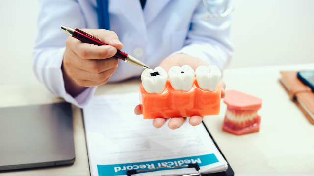
A resin restoration is usually enough when decay is small and the tooth's walls and cusps are intact. Larger damage points to an inlay, onlay, or crown.
Learn More
Inlay Onlay Dental Restoration vs Crown: How to Choose
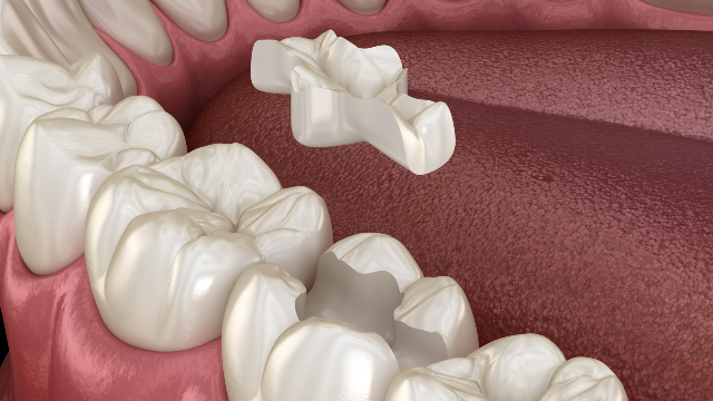
An inlay or onlay restores a damaged tooth without full coverage. Published data show partial-coverage survival close to crowns, so choose by tooth left.
Learn More
Minish Dental Treatment: How It Preserves Tooth Structure
Minish dental treatment is a biomimetic restoration that rebuilds damaged teeth with precision-made blocks while removing as little enamel as possible.
Learn More